Tuesday, March 16, 2021

Mobile Radiography - Advanced Technology of Imaging

 Radiography is an imaging technique that used gamma rays, X-rays and other electromagnetic radiation to image the internal parts of a human body. Mobile radiography is basically used for bedside radiography examinations of patients who cannot be transported to the medical imaging room.

Download Sample PDF Brochure Of Mobile Radiography
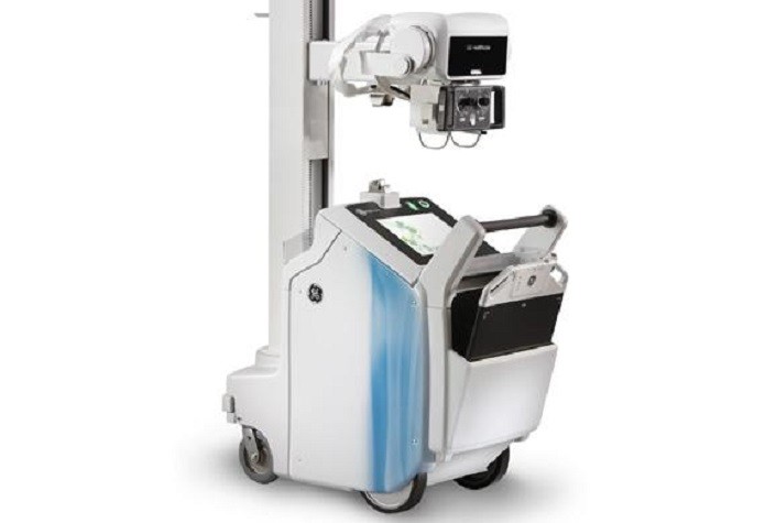
Mobile X-ray systems are used throughout the hospital from in-patient, to NICU, Operating Room, and Emergency Room imaging. The need for point of care, fast, reliable, and high IQ imaging is growing as technology shifts from analog / CR cassettes to wireless flat panel. Worldwide, Hospitals and Imaging centers are witnessing a high volume of patients requiring quick and efficient imaging of COVID-19 suspect cases.

Mobile X-ray equipment are being used as an important triaging and screening tool for determining the condition of the lung before being directed to advanced imaging techniques. Even after medical intervention, when the patient is in ICUs or Ambulatory care, Mobile X-rays may be used for monitoring the condition of lungs through regular chest X-rays. A Mobile X-Ray equipment is easy to setup, quick to move to the patient, easy to park next to bedside, and easy to disinfect and prepare for the next exam. The Mobile Radiography Systems market is anticipated to grow in the forecast, owing to the factors such as increasing incidence of chronic disease such as diabetes, cardiovascular disease etc., rising adoption of technologically advanced health care devices, rising geriatric population, advancements in healthcare, and growing number of medical imaging procedures.

Access Full PDF

Wednesday, March 3, 2021

Introduction to Neuroscience: Understanding the Brain

Neuroscience (or neurobiology) is the scientific study of the nervous system. It is a multidisciplinary science that combines physiology, anatomy, molecular biology, developmental biology, cytology, computer science, mathematical modeling, and psychology to understand the fundamental and emergent properties of neurons and neural circuits. The understanding of the biological basis of learning, memory, behavior, perception, and consciousness has been described by Eric Kandel as the "ultimate challenge" of the biological sciences. Tools such as MRI scans and computerized 3-D models are used to perform tests for some common conditions including Down syndrome, Parkinson's disease, brain tumors, effects of stroke such as, language loss and many others.

Download Sample Copy Of Neuroscience

Humans have an estimated hundred billion neurons, or brain cells, each with about a thousand connections to other cells. One of the great challenges of modern neuroscience is to map out all the networks of cell-to-cell communication—the brain circuits that process all thoughts, feelings, and behaviors. The resulting picture, emerging bit by bit, is known as "the connectome." The ability of the brain to elaborate new connections and neuronal circuits—neuroplasticity—underlies all learning.

A rapidly expanding discipline, neuroscience findings have grown by leaps and bounds over the past half-century. More work, however, will always be needed to fully understand the neural roots of human behavior, consciousness, and memory.

Thursday, February 25, 2021

Control of Gene Expression By Regulation

 Gene expression is a technique through which genetic instruction are used for synthesizing gene products. This technique enables scientists and researchers to reach at the molecular level of each gene. Proteins are generally synthesized with the help of gene expression which further perform the function of components such as proteins, enzymes as well as receptors. Process of gene expression involves of two stages, transcription and translation. The techniques used for monitoring the gene expression levels include, northern blot analysis, RNA protection assay, and microarrays among others.

Download Sample Copy Of Gene Expression


Control of gene expression is essential to all organisms. In bacteria, it allows the cell to take advantage of changing environmental conditions. In multicellular organisms, it is critical for directing development and maintaining homeostasis. Gene regulation can occur at three possible places in the production of an active gene product. First, the transcription of the gene can be regulated. This is known as transcriptional regulation. When the gene is transcribed and how much it is transcribed influences the amount of gene product that is made. Second, if the gene encodes a protein, it can be regulated at the translational level. This is known as translational regulation. How often the mRNA is translated influences the amount of gene product that is made. Third, gene products can be regulated after they are completely synthesized by either post-transcriptional or post-translational regulation mechanisms. Both RNA and protein can be regulated by degradation to control how much active gene product is present.

Access Full PDF


Monday, February 22, 2021

A Brief Review of Brain Monitoring Technology: Current Status and Future Directions

Brain monitoring devices are used to monitor brain activities of the patients during their conscious and unconscious conditions. Some of the monitors are used to measure the oxygen level and provides accurate, consistent measurements of oxygen in tissue.

Brain monitoring is important in critical clinical scenarios where the extent or evolution of neurologic injury is unknown. Common situations include the comatose state after cardiac arrest, poor grade aneurysmal subarachnoid hemorrhage (aSAH), and severe traumatic brain injury (TBI) et al. The extent of neurologic injury and injury progression are crucial in determining prognosis and guiding intensive care strategies to ameliorate the neurologic injury.

Download Sample PDF of Brain monitoring

Brain injury occurs either at the time of a direct insult or subsequently due to changes in the physical and biochemical environment. The ability to measure these changes reliably is of paramount importance in order to tailor the treatment to each individual patient and therefore prevent the onset of secondary brain injury.


Multimodal brain monitoring can be grouped into three categories:

(1)direct signals which are monitored invasively (e.g., intracranial pressure (ICP), tissue oxygenation, microdialysis, parenchymal blood flow, etc.);

(2)variables which may be monitored noninvasively (e.g., transcranial Doppler (TCD) or near infrared spectroscopy (NIRS));

(3)variables describing brain pathophysiology which are not monitored directly but are calculated at the bedside by dedicated computer software. The simplest example is the cerebral perfusion pressure (CPP), which is the difference between the mean arterial blood pressure (MAP) and the ICP , and therefore it is a calculated variable. More sophisticated examples include various indices of vascular reactivity or cerebral autoregulation, brain compensatory reserve, vascular resistances, and brain compartmental compliances.

Optogenetics—Possible Future Monitoring Tool?

Optogenetics is an interesting area of research relatively in its infancy but may have potential clinical applications in brain monitoring. In its simplest permutation, optogenetics involves optically recording changes in membrane potential. Regarding the central nervous system, changes in membrane potential is how action potentials spread down an axon, across a synapse, and activate second order neurons. The ability to see neuronal signal transmission in action implicates that functional circuitry can be characterized. The functional assessment possible through optogenetics is an advantage over current brain monitoring techniques.

Access Full PDF 

Thursday, February 11, 2021

Human Genetics : Basic Concepts & Application

Human genetics includes study of the inheritance in humans. Human genetics is broad sector which include various sub-sectors such as cytogenetics, molecular genetics, biochemistry, linical genetics, and genetic counseling and others. These sectors helps in understanding the concepts of gene structure and organization, expression of gene, detection of mutation and its analysis, linkage analysis and genetic mapping, and physical mapping among others. The study of human genetics allows to understand genetically of complexes in a diseases and epistatic interactions such as ethical, legal and social issues.

Get Sample PDF Of Human Genetics


• The genetic information of an individual is contained in 23 pairs of chromosomes.

Every human cell contains the 23 pair of chromosomes.

• One pair is called sex chromosomes

 Male: XY

 Female: XX

• Other 22 pairs of homologous chromosomes are called autosomes.

• The autosome chromosome pairs are called homologous pair. Two chromosomes in the same pair are called homologous chromosomes. 

Human Genome

• The totality of DNA characteristic of all the 23 pairs of chromosomes.

⎯ The human genome has about 3x10⁹ bps in length.

⎯ 97% of the human genome is non-coding regions called introns. 3% is responsible for controlling the human genetic behavior. The coding region is called extron.

⎯ There are totally about 40,000 genes, over 5000 have been identified. There are much more left

⎯ Human Genome Project is to identified the DNA sequence (every bp) of human genome ( only a few individuals)

⎯ For human being, most of the place in human genome are the same. Only a very small part is different among different individuals. 

Practical Application of Human Genetics Recognizing that the goal of most introductory science courses is to better inform future voters and consumers, the author provides practical application of the content to students’ lives. Topics of particular interest to students include:

■ The role that genes play in disease susceptibility, physical characteristics, body weight, and behaviors, with an eye toward the dangers of genetic determinism

■ Biotechnologies, including genetic testing, gene therapy, stem cell therapy, gene expression profiling, genomewide association studies, and personalized medicine

■ Ethical concerns that arise from the interface of genetic information and privacy, such as inf such as infidelity testing, ancestry testing, and direct-to-consumer genetic testing 

Access Full PDF

Thursday, February 4, 2021

Exercises to Help Improve Brain Health | Strengthen Your Mind and Thrive

Things you can do to help maintain your brain health

Brain fitness is having brain-based emotional, cognitive, and self-regulation capacities needed to succeed in one's environment. Brain fitness refers to different techniques and strategies such as cognitive training to keep the brain 'in shape' by being engaged in mental exercises that target and engage the senses, memory, and attention. For all ages, as well as for people suffering from ADHD, learning disorders, memory loss, and even brain injury, keeping the brain' fit' is recognised as helpful and important.

Get Sample PDF Of Brain Fitnees


Neuroscience is discovering new, effective ways to develop our brains for enhanced mental strength and performance. What we are learning about neuroplasticity means that, at any age, through training, we can change our brains to become even more mentally strong, healthy, and fit. Much as we need specific physical exercises and movement to build bodily strength, agility, and resilience; we can also benefit from exercises — or practices — to strengthen our minds, mental performance, and well-being. We can tap this immense potential to experience greater courage, confidence, composure, clarity, and creativity, the 5 Cs of mental strength we need to cultivate happier, healthier, and more rewarding lives.

Brain fitness has quickly become a mainstream aspiration among baby boomers and elders, primarily in North America. It has fueled a growing interest in brain fitness classes, brain fitness centers, and brain fitness programs, along with attendant opportunities and challenges. An increasing number of adults want useful tools to protect cognitive health and performance—not necessarily to reverse aging—and what they are finding is an expanding and noisy marketplace where they (and also professionals) need to carefully evaluate their own needs and the available options. The recent discovery that experience can change brain structure and function at any age has inspired a range of health, education, and productivity applications whose value and limitations we are only starting to grasp. If you can envision the array of equipment available to train different muscles in a typical modern health club, you can anticipate the value—and perhaps the limitations—of having an expanding toolkit to measure and enhance cognition and mental wellness. The burgeoning brain fitness industry needs to define and refine itself, to mature, before it can be as established as today’s physical fitness industry.

Access Full PDF

Tuesday, January 26, 2021

Guidelines for Gene Therapy (Gene-modified) Product Development

Genetic modification therapy incorporates a functional gene into a cell-based therapy. The genetic modification generally occurs outside the body, but the resulting genetic change to the patient's DNA is permanent. The active payload comprise of the gene encoding for production of the therapeutic protein and gene controls that regulates production of the therapeutic gene. The vector, which may be either non-viral or viral, delivers the gene and gene control payload to the cells which are to be genetically modified. The most common cell types include T cells, and HSCs. Genetic modification has brought a milestone into medicine. Genetic modification is very crucial in treating severe diseases.

Download Sample PDF Of Genetic Modification Therapy


Gene therapy gathered pace over the past couple of decades due to the rapid discovery of large numbers of genes which are mutated in a variety of diseases. Such diseases may be inherited diseases caused by single gene mutations or polygenic, complex disorders where many genes are mutated or their expression is affected. Gene therapy, can treat inherited diseases by providing a fully functional, “wild-type” copy of the gene which can recapitulate the functions required in the cell. However, in complex disorders such as cancers, gene therapy is often synthetic in strategy, wherein novel genes, such as suicide genes or tumor suppressors may be introduced into the tumor to selectively kill the cancer cells, or reprogram immune cells in the body to target the cancerous tissue.

Progress in the therapeutic efficacy of gene-modified cell therapies across a spectrum of both preclinical models and clinical trials has renewed optimism in these much-anticipated next generation of “drugs.” Advances in genetic engineering now make development of these therapeutic cells more accessible to scientists—allowing for their wider adoption, more fine-tuned development, and ultimately, more diverse application. This special Cytotherapy Issue highlights the rapidly expanding field of gene-modified cells for therapy of a variety of malignant and non-malignant disorders.

Access Full PDF

Wednesday, January 20, 2021

A Guide to Genetic Tests that are used to Examine Manygenes at the Same time - Genetic Diagnosis, Risk Prediction

Gene panel testing is known as an option for genetic testing and counseling associated to cancer risks. The Gene panel test is used to perform to analyze the multiple genes at once for the cancer-associated mutations. The test is capable to examine a several number of genes that can provide information related to the cancer and provide a secure diagnostics to help to prevent or stop the cancer to be spread.

 What are genes?

Our bodies are made up of millions of cells. Most of those cells contain a complete set of genes. Genes act like a set of instructions, controlling our growth and how our bodies work. They are also responsible for many of our characteristics, such as our eye colour, blood type or height. We have thousands of genes. We each inherit two copies of most genes, one copy from our mother and one copy from our father. That is why we often have similar characteristics to both of our parents. The genes are composed of specific sections of DNA1 which are a bit like the instruction manual inside the gene.

Download Sample PDF Of Gene Panel


Gene sequencing panels are a powerful diagnostic tool for many clinical presentations associated with genetic disorders. Advances in DNA sequencing technology have made gene panels more economical, flexible, and efficient. Because the genes included on gene panels vary widely between laboratories in gene content (e.g., number, reason for inclusion, evidence level for gene–disease association) and technical completeness (e.g., depth of coverage), standards that address technical and clinical aspects of gene panels are needed. This document serves as a technical standard for laboratories designing, offering, and reporting gene panel testing.

Although these principles can apply to multiple indications for genetic testing, the primary focus is on diagnostic gene panels (as opposed to carrier screening or predictive testing) with emphasis on technical considerations for the specific genes being tested.

PANEL DESIGN CONSIDERATIONS

Phenotype-directed diagnostic gene panels Due to the sequencing capacity of current technologies, hundreds to thousands of genes can now be delved simultaneously to determine the cause of genetic disorders.

Examples of several phenotype-directed panels and subpanels are listed in Table 1. The goal of a diagnostic gene panel is to maximize clinical sensitivity and minimize the clinical burden from analysis of inappropriate or unnecessary genes that may result in variants of uncertain clinical significance (VUS). Patients may have limited opportunity for serial genetic tests, arguing for casting a wide diagnostic net; however, long lists of

(VUS), a possible outcome from testing large numbers of genes, can complicate medical management and cause unnecessary patient anxiety. When a panel is well-designed, it will

● Be cost-effective for a particular clinical indication.

● Maximize clinical sensitivity by, to the extent possible, including all GADs associated with a disorder, therebyallowing disorders with clinical heterogeneity and overlapping features to be molecularly diagnosed.

● Include GUSs with limited but emerging evidence ifadditional criteria are met (see example in “Clinicalsensitivity,” “Gene considerations,” and “Reporting”).

● Maximize clinical specificity by limiting or excludingGUSs, thereby minimizing detection of VUS.

● Employ auxiliary assays for genes/regions that cannot beinterrogated with current sequencing technology tomaximize the clinical utility.

Access Full PDF


Friday, January 8, 2021

Neuronavigation Systems: Concept, Techniques, Clinical Applications and Potential Pitfalls

Neuro-navigation, also called as frameless stereotactic surgery is the technique which involves performance of real-time intraoperative guidance during spinal or brain injuries. This increases safety and accuracy during neurosurgery. Neuro-navigation systems help to guide the surgeon to the surgical targets without the need for external frames. These systems are used majorly in brain surgeries which helps to limit the size of skull opening or craniotomy and remove brain lesions such as tumors or other tissue masses.

Download Sample PDF Of Neuronavigation Systems


There are significant technical issues that need to be addressed and understood in the use of such equipment in order to minimize inaccuracy. The most significant limitation of neuronavigation is brain shift. Brain shift can be minimized by selecting the site of the craniotomy uppermost in the operative field and by limited use of retraction, however, it is inevitable and can lead to limited application of the neuronavigation system over time during surgery. The loss of registration that can occur due to inadvertent intraoperative manipulation of the reference frame or loss of fixation of the head, can be retrieved by the registration of intraoperative bony landmarks at the margins of the craniotomy.

Localization and delineation of extent of lesions are critical for safe maximal resection of brain and spinal cord tumors. Neuronavigation systems have been developed for image-guided neurosurgery to aid in the accurate resection of brain tumors. Basic principles of navigated surgery are to see the tip of a pointer in an image space. A relationship between the device space and the image space has to be established. This operation is called registration or calibration of the navigation device. Basically, a transformation matrix (T) has to be calculated to map the coordinates of any point between the image and the device spaces. The aim of transformation matrix is to create a linkage between digital image data and anatomical structure, and therefore, to provide increasing 3-D orientation. Today's navigation systems provide approximately 2mm accuracy.

Stereoscopic navigation-controlled display of preoperative MRI and intraoperative 3D ultrasound is a new technology for minimally invasive image-guided surgery approaches in planning and guiding neurosurgery. Interactive stereoscopic visualization improves perception and enhances the ability to understand complex 3D anatomy. In 1947, Spiegel and Wycis performed the first stereotactic thalamotomy in humans, using the commissura posterior or pineal body as an internal individual reference system. Functional operations with similar frames and techniques were introduced by Talairach in Paris in 1949, by Riechert in Freiburg, Germany in 1952, and by Leksell in Stockholm in 1949 for the treatment of extrapyramidal movement disorders, intractable pain, epilepsy, and psychiatric disorders.

Advantages and disadvantages

There are some concerns about navigation systems including time consuming calculation and registration, restriction of space and view inside the operating field,and so on. Nevertheless, there are many advantages that can be helpful in the process of operation

Access Full PDF

Wednesday, December 23, 2020

Building Your Digital DNA - Trend Of Digital Transformation (Digital Genome)

Digital genome is a complete digital set of genetic material that present in an organism or a cell. Digital genome technology deals with genes and their functions to find the causes behind the chronic diseases and also to fix them. The technology is associated with the advancements that help to make healthcare more personal and more effective for the treatment. Moreover, the digital genome is an easier way of gathering  information about the chronic disease. The technology is used by the professionals to get a closer look of genetic composed diseases, such as cancer. Digital genome acts as a supporter that enables instant access to trait combinations to solve apparently endless custom queries.

Download Sample PDF Of Digital Genome


New genome engineering technologies, like CRISPR-Cas9, are revolutionising how we think about genetics. The ability to make targeted changes to a living cell’s genome has huge implications in fields like precision health, agriculture and biosecurity.

But genomes are big, and everyone's is different. How do you design a tailored approach for a specific genome? How do you know where to target amongst the billion bases in a human genome?

As digital information continues to accumulate, higher density and longer-term storage solutions are necessary. DNA has many potential advantages as a medium for immutable, high latency information storage needs. For example, DNA storage is very dense. At theoretical maximum, DNA can encode two bits per nucleotide (nt) or 455 exabytes per gram of ssDNA. Unlike most digital storage media, DNA storage is not restricted to a planar layer, and is often readable despite degradation in non-ideal conditions over millennia. Finally, DNA’s essential biological role provides access to natural reading and writing enzymes and ensures that DNA will remain a readable standard for the foreseeable future. 

Genome editing is a powerful new tool for making precise alterations to an organism’s genetic material. Recent scientific advances have made genome editing more efficient, precise, and flexible than ever before. These advances have spurred an explosion of interest from around the globe in the possible ways in which genome editing can improve human health. The speed at which these technologies are being developed and applied has led many policymakers and stakeholders to express concern about whether appropriate systems are in place to govern these technologies and how and when the public should be engaged in these decisions.

Access Full PDF

Saturday, December 19, 2020

CRISPR Technology- Latest Advances and Upcoming Prospects for Genome Editing

CRISPR (Clustered Regularly Interspaced Short Palindromic Repeats) technology is a simple but powerful tool for genome editing. This tool enables life science researchers to easily edit DNA sequences and modify gene function. It has many potential applications include correcting genetic defects, treating and preventing the spread of diseases and improving crops. By delivering the CRISPR enzyme Cas9 nuclease coupled with synthetic guide RNA (gRNA) into a cell, the cell's genome can be cut at a desired location, that allows existing genes to be removed or add new ones.

Download Sample PDF of CRISPR Technology


Increasing usage of CRISPR systems in microbiology, growing government and private investments on research and development of genome editing, rising prevalence of genetic disorders, and increases application of CRISPR/Cas9 technology to improve crop production drives the global CRISPR technology market. However, ethical issues associated with CRISPR and lack of skilled personnel restrain the global CRISPR technology market over the forecast period.

CRISPR is igniting a revolution. A relatively recent discovery in the timeline of biotechnology, CRISPR is quickly becoming a standard and flexible laboratory tool, and it is well on its way to permeating a large variety of applications. Researchers are deploying CRISPR across a wide range of life science disciplines, from agriculture and medicine to biofuels and industrial fermentation.

Genome editing (also called gene editing) is a group of technologies that give scientists the ability to change an organism's DNA. These technologies allow genetic material to be added, removed, or altered at particular locations in the genome. Several approaches to genome editing have been developed. A recent one is known as CRISPR-Cas9, which is short for clustered regularly interspaced short palindromic repeats and CRISPR-associated protein 9. The CRISPR-Cas9 system has generated a lot of excitement in the scientific community because it is faster, cheaper, more accurate, and more efficient than other existing genome editing methods.

CRISPR-Cas9 was adapted from a naturally occurring genome editing system in bacteria. The bacteria capture snippets of DNA from invading viruses and use them to create DNA segments known as CRISPR arrays. The CRISPR arrays allow the bacteria to "remember" the viruses (or closely related ones). If the viruses attack again, the bacteria produce RNA segments from the CRISPR arrays to target the viruses' DNA. The bacteria then use Cas9 or a similar enzyme to cut the DNA apart, which disables the virus.

Access Full PDF

Wednesday, December 16, 2020

New Techniques in Digital Radiology : Improves Image Quality & Many More

Advances in digital technology allowed the development of full digital X-ray detectors that are currently available for projection radiology. Digital radiography is a method of radiography that uses x-ray-sensitive plates to capture data during the patient examination directly and immediately transferring it to a computer system without the use of a standard cassette. The computed radiography process is to use a phosphor imaging plate to create a digital image. CR uses a cassette-based system like analog film. The DR technology transfer automatically the images to a computer.

Download Sample PDF Brochure!


Digital radiography is a method of radiography that uses x-ray-sensitive plates to capture data during the patient examination directly and immediately transferring it to a computer system without the use of a standard cassette. The computed radiography process is to use a phosphor imaging plate to create a digital image. CR uses a cassette-based system like analog film. The DR technology transfer automatically the images to a computer. Digital radiography has revolutionized and streamlined endodontic diagnosis and treatment. There are several advantages of digital x-ray imaging over analog film imaging that can benefit the clinician: reduced time, reduced radiation, ability to take multiple exposures without repositioning the sensor, storage and maintenance of the images, and electronic transmission of images. Digital radiography presents numerous advantages: a big dynamics bounded to an mostly linear response; an interactive parameter setting the examinations devices; aquasi-immediate availability of the image; operations of post-treatment on the images in order to improve their quality; the availability and the sharing of the images on local or distant stations. Today, the greater needs in flexibility and "real time", as well for the acquisition of the images as for the communications, the sharing of information, the processing and the computing exploitation of the big data bases of images, as well as the more and more frequent exploitation of the temporal dimension (movies-radiography) make the use of one of these new systems of digital radiography almost indispensable. The emergence of these technologies was allowed by progress of electronics and computing science and needs in instrumentation evolved at the same time as the increase of the performances of the computing simulation tools. The either optoelectronics or semiconductor new sensors achieve performances which can on certain points exceed those of the film systems: • high power of ruling allowing a high threshold of detection, • noise only limited by the statistics of detected photons X, • dynamics above 1000, • linearity on all the range of signal, • speed of evacuation of the data • frame rate…

Access Full PDF

Sunday, December 13, 2020

Recent Developments of Reporter Gene Assays - Methods and Protocols (with Appilications)

Reporter Genes are genes whose products can be readily assayed consecutive to transfection and can be used as markers for screening successfully transfected cells, for studying regulation of gene expression, or serve as controls for standardizing transfection efficiencies.

Gene reporter systems play a key role in gene expression and regulation studies. This review describes the ideal reporter systems, including reporter expression vector design. It summarizes the many uses of genetic reporters and outlines the currently available and commonly used reporter systems. Each system is described in terms of the reporter gene, the protein it encodes, and the assays available for detecting presence of the reporter. In addition, each reporter system is analyzed in terms of its recommended uses, advantages, and limitations.

Download Sample PDF Of Brochure, Click Here!

A gene consists of two main functional elements: The first consists of a so-called coding DNA sequence that provides information about the protein produced. Second, a specific promoter sequence linked to the coding region that regulates the transcription of the gene. The promoter serves to activate or suppress the expression of the gene as required.

The main purpose of the reporter gene assay is to investigate the promoter of a gene of interest, i.e. the regulation of its expression. This can be done by linking the promoter of interest to an easily detectable gene, such as the gene for firefly luciferase, which catalyses a reaction that produces light. 

Usually, the cells are then exposed to different factors or conditions, or changes can be made in the order of the reporter, the effect of which can be easily tracked by measuring changes in light emission.

EXAMPLES OF REPORTER GENES

Common reporter genes are β-galactosidase, β-glucuronidase and luciferase. Various detection methods (see below) are used to measure expressed reporter gene protein. These include luminescence, absorbance and fluorescence.

Assays based on luminescence are very popular for several reasons:

  • they have a high sensitivity (between 10 and 10,000 times higher than methods based on absorption or fluorescence, depending on the specific assay and reporter used)
  • most cell types do not have endogenous luciferase activity
  • luminescence assays have a large dynamic range
  • they are quick to perform
  • their costs are relatively low 

Friday, December 11, 2020

Benefits & Importance of Wireless Brain Sensors : The Human Machine Interface that Control Your Thinking

Over the past several decades, the use of electronics in a wearable form to monitor body signals and the surrounding environment has led to major developments in healthcare and environmental sensors. According to the World Health Organization (WHO), brain injury is the main source of death and inability around the world. It is evaluated that roughly 10 million individuals will be influenced by traumatic brain injury (TBI) by 2020.

Download Sample PDF Brochure, Click Here!


Wireless implantable sensors brings the promising prospects for the treatment of intracranial diseases. To achieve the transcranial wireless links for data transmission between the implanted sensor and the off-body receiver, different approaches have been proposed in the literature.In this summary paper, we will introduce and briefly analyze the state of the art and then introduce two implantable battery-free systems for brain care application from our current research. Wireless brain sensors are detects the intracranial pressure, monitor the brain temperature and record brain signaling through the form of brain waves. Wireless brain sensors are mostly used for patients with conditions such as sleep disorders, Parkinson's disease, traumatic brain injury, dementia and other brain related conditions. These sensors assists in monitoring the neurological fluctuations and help to improve cognitive functionalities. These sensors operates wirelessly, and can be easily accessible through wireless connectivity with the help of smart phone, tablets or computers and making the device more cost-efficient.

Wireless brain sensors are devices that help monitoring the temperature, detecting the intracranial pressure, and record brain signaling in the form of brain waves. The essential aim of this wireless brain sensor is of securing the person from emergency situations. The devices are primarily used for patients experiencing conditions such as sleep disorders, traumatic brain injury, dementia, Parkinson’s disease, and other neurological conditions. These devices aid in observing and monitoring the neurological deviations and provide support for improving the cognitive functionalities. Accessibility of these sensors is easy from a remote area through wireless connectivity and be integrated with smart phones, tablets and computers, consequently be monitored intermittently from a homecare environment, making the device more cost-efficient.


Wednesday, December 9, 2020

Genomics and Proteomics : Concepts and Perspectives, Applications

Proteomics is the study of the complete protein complement of a cell, tissue, or organism under a precise, defined set of conditions. It is depended on technological and instrumental developments. These developments comprised of advances in mass spectrometry (MS) technology, protein fractionation techniques, bioinformatics, etc. Proteomics depend on three basic technological bases that include a method to fractionate compound protein or peptide mixtures, MS to acquire the data necessary to classify individual proteins and bioinformatics to analyze and assemble the MS data.

The word “proteome” represents the complete protein pool of an organism encoded by the genome. In broader term, Proteomics, is defined as the total protein content of a cell or that of an organism. Proteomics helps in understanding of alteration in protein expression during different stages of life cycle or under stress condition. Likewise, Proteomics helps in understanding the structure and function of different proteins as well as protein-protein interactions of an organism.

Download Sample PDF Brochure, Click Here!

Types of Proteomics

Expression proteomics: The quantitative study of protein expression between samples thatdiffer by some variable is known as expression proteomics. Using this approach, protein expression of the entire proteome or of subproteomes between samples can be compared. This could be useful in identification of disease specific proteins.

Structural proteomics: Unlike comparing the same cell or tissue in normal and diseased state inexpression proteomics, structural proteomics are directed to map out the structure and nature of protein complexes present specifically in a particular cellular organelle. The aim is to identify all proteins present in a complex and to characterize all protein-protein interactions occurring between these proteins.

Functional proteomics: Functional proteomics is a very broad term for many specific, directedproteomics approaches. It can be defined as the use of proteomics methods to analyze the properties of molecular networks involved in a living cell. One of the major objectives is to identify molecules that participate in these networks.

Genes and Proteins

Number of genes vs Number of proteins

Analysis of mRNA does not provide a direct reflection of the protein content in the cell. One reason is that mRNA and protein expression levels do not always correlate. mRNA could undergo various post-transcriptional modifications like, polyadenylation and mRNA editing. Some of these modifications could lead to the generation of various protein forms from a single gene. Subsequently, translational regulation of mRNA could take place. Proteins, after synthesis, could undergo post-translational modifications. It is estimated that proteins could undergo as many as 200 different types of these modifications.

Access Full PDF

Monday, December 7, 2020

Gene Expression and Regulation

Gene expression is a technique through which genetic instruction are used for synthesizing gene products. This technique enables scientists and researchers to reach at the molecular level of each gene. Proteins are generally synthesized with the help of gene expression which further perform the function of components such as proteins, enzymes as well as receptors. Process of gene expression involves of two stages, transcription and translation. The techniques used for monitoring the gene expression levels include, northern blot analysis, RNA protection assay, and microarrays among others.

The decreasing cost of sequencing techniques and availability of gene expression databases are expected to fuel the growth of market in the coming years. Increase in the development of biotechnology sector in various developing economies coupled with the funding offered by the government bodies are anticipated to offer growth opportunities in the market during the forecast period.

Download Sample PDF Brochure, Click Here

What is Gene Expression?

Gene expression is the process in which DNA directs the synthesis of functional products, such as proteins. Cells can regulate gene expression at various stages. It allows organisms to generate different cell types and enables cells to adapt to internal and external factors.

  • Gene expression is a tightly regulated process that allows a cell to respond to its changing environment.
  • It acts as both an on/off switch to control when proteins are made and also a volume control that increases or decreases the amount of proteins made.
  • There are two key steps involved in making a protein, transcription and translation.

Genetic Information Flows from DNA to RNA to Protein

A gene is a stretch of DNA that serves as the blueprint for functional RNAs and proteins. Since DNA is made up of nucleotides and proteins consist of amino acids, a mediator is required to convert the information that is encoded in DNA into proteins. This mediator is the messenger RNA (mRNA). mRNA copies the blueprint from DNA by a process called transcription. In eukaryotes, transcription takes place in the nucleus by complementary base-pairing with the DNA template. The mRNA is then processed and transported into the cytoplasm where it serves as a template for protein synthesis during translation. In prokaryotes, which lack a nucleus, the processes of transcription and translation occur at the same location and almost simultaneously since the newly-formed mRNA is susceptible to rapid degradation.

Access Full PDF

Wednesday, December 2, 2020

Role of Epigenetics in Biology - Advancing Diagnostics and Accelerating Precision Medicine

 Epigenetics is a study of change in gene expression in a phenotype without changing the genotype. Epigenetics drugs and diagnostics are used while screening early stage cancer and other abnormal changes in skin cells, liver cells, brain cells and others.

There are pronounced interindividual variations in drug metabolism, drug responses, and the incidence of adverse drug reactions. To a certain extent such variability can be explained by genetic factors, but epigenetic modifications, which are relatively scarcely described so far, also contribute. It is known that a novel class of drugs termed epidrugs intervene in the epigenetic control of gene expression, and many of these are now in clinical trials for disease treatment. In addition, disease prognosis and drug treatment success can be monitored using epigenetic biomarkers. Here we review these novel aspects in pharmacology and address intriguing future opportunities for gene-specific epigenetic editing. 

Download Sample PDF Brochure Of Study, Click Here!


Epigenetic mechanisms can influence the gene activity at the transcriptional and post-transcriptional levels and/or at the translation level and post-translational modifications. Such epigenetic mechanisms with a potentially vast spectrum of consequences could result in more varieties of cell differentiations, morphogenesis, variability, and adaptability of an organism, which can be affected by both genetic and environmental factors. Therefore, the field of epigenetics covers the modifications of DNA, DNA-binding proteins, and histones, which are important in making changes in chromatin structure without any change in the nucleotide sequence of a given DNA. Also, some of these alterations could be transferred between generations. The advent of the epigenomic tools over the past few years has brought forth a more holistic view of the interplay between the genome and a very active epigenome, forming an causal link between the underlying genome, the regulatory epigenome, and the functional consequences stemming from perturbations in both. Importantly, these tools are being leveraged to uncover mechanisms of complex diseases. Such integrative approaches may provide insights into the causal regulatory mechanisms of disease for purposes of early-stage detection as well as therapeutic development.

The exciting and rapidly evolving area of epigenetics is advancing the possibilities for medicine well beyond the realms of the genome, bringing a new generation of diagnostic and therapeutic innovations that offer hope for many patients living with life-threatening diseases.

Access Full PDF Brochure

Wednesday, November 25, 2020

What Is a Brain Pacemakers? How Pacemakers Treat Parkinson’s Disease?

 Electricity Restoring Function & Replacing Drugs

A brain pacemaker is a medical device implanted into the brain to stimulate the nervous tissues with electric signals. These pacemakers are being used widely to provide treatment to the patients having neurological disorders such as Parkinson's disease, epilepsy, and others. Other than giving stimulation to the brain, pacemakers also play an essential role in stimulating the spinal cord. Brain pacemakers have been found to offer a safe and effective procedure that provides symptomatic relief to patients. Doctor puts the pacemaker under the skin on your chest, just under your collarbone. It’s hooked up to your heart with tiny wires.

Download Sample PDF Brochure Of Study, Click Here


Doctors know that deep brain stimulation works as a therapy for Parkinson’s disease. But they’re still trying to figure out why and how. A new study sheds some light on the mechanism of action, suggesting that deep brain stimulation disrupts a pattern of excessively synchronized activity in the brain. In DBS, an implanted device sends tiny jolts of electricity through neurons, acting somewhat like a brain pacemaker. The technique is widely accepted as a treatment for Parkinson’s and other movement disorders; more than 100,000 patients have received implants that help control their tremors, rigidity, and other kinetic symptoms.

How does Pacemakers work?
• A pacemaker uses batteries to send electric signals to your heart to help it pump the right way.
• The pacemaker is connected to your heart by one or more wires. Tiny electric charges that you can’t feel move through the wire to your heart.
• Pacemakers work only when needed. They go on when your heartbeat is too slow, too fast or irregular.

The Brain Pacemaker has already revolutionized the treatment of movement disorders and in the future will completely alter the management of other disabling conditions. Our AIMS are to learn exactly how electrical stimulation normalizes brain function, to develop better technology for the device and other neural prosthetics, test these systems for new conditions, and thus improve patient’s quality of life.

Access Full PDF Brochure!

Friday, November 20, 2020

Quality Systems For Medical Imaging - Essentials for Physicians

Medical imaging is a process to examine the anatomical & physiological conditions of the human body for clinical analysis or diagnosis. It includes different processes to create image of internal organs & tissues for use in disease monitoring and treatment purposes. Medical imaging comprises three types of imaging- radiology imaging, nuclear imaging and optical imaging. Medical imaging seeks to reveal internal structures hidden by the skin and bones, as well as to diagnose and treat disease. Medical imaging also establishes a database of normal anatomy and physiology to make it possible to identify abnormalities. Although imaging of removed organs and tissues can be performed for medical reasons, such procedures are usually considered part of pathology instead of medical imaging.

Download Sample PDF Brochure Of Study


As a discipline and in its widest sense, it is part of biological imaging and incorporates radiology, which uses the imaging technologies of X-ray radiography, magnetic resonance imaging, ultrasound, endoscopy, elastography, tactile imaging, thermography, medical photography, and nuclear medicine functional imaging techniques as positron emission tomography (PET) and single-photon emission computed tomography (SPECT). Measurement and recording techniques that are not primarily designed to produce images, such as electroencephalography (EEG), magnetoencephalography (MEG), electrocardiography (ECG), and others, represent other technologies that produce data susceptible to representation as a parameter graph vs. time or maps that contain data about the measurement locations. In a limited comparison, these technologies can be considered forms of medical imaging in another discipline.

Medical imaging is often perceived to designate the set of techniques that noninvasively produce images of the internal aspect of the body. In this restricted sense, medical imaging can be seen as the solution of mathematical inverse problems. This means that cause (the properties of living tissue) is inferred from effect (the observed signal). In the case of medical ultrasound, the probe consists of ultrasonic pressure waves and echoes that go inside the tissue to show the internal structure. In the case of projectional radiography, the probe uses X-ray radiation, which is absorbed at different rates by different tissue types such as bone, muscle, and fat.

For More info in detail, Click here

Tuesday, November 17, 2020

Progress in Neuroengineering for Brain Repair | Future of Brain Implants, Potentially Safer way to Study

Intracerebral implants make use of drug-impregnated polymers that allow controlled release of the drug at the desired site in the brain over an extended period of time.

Brain implants are neural implants that are used to stimulate the parts & structures of the nervous system. These implants are technical systems that communicate with the nervous system and help to enhance senses, physical movement, and memory after a stroke or other head injuries. Deep brain stimulation and spinal cord stimulation are used to treat depression, obsessive-compulsive disorder and epilepsy, among other neural disorders.

Download Sample PDF Brochure of Brain Implants


This includes sensory substitution , e.g. in vision . Brain implants involve creating interfaces between neural systems and computer chips , popularly called brain-machine interfaces .Other brain implants are used in animal experiments simply to record brain activity for scientific reasons. Some brain implants involve creating interfaces between neural systems and computer chips. This work is part of a wider research field called brain-computer interfaces. (Brain-computer interface research also includes technology such as EEG arrays that allow interface between mind and machine but do not require direct implantation of a device.)

Neural implants such as deep brain stimulation and Vagus nerve stimulation are increasingly becoming routine for patients with Parkinson's disease and clinical depression, respectively. Next-generation implants may someday treat diseases of the brain itself, particularly those of aging. Such ailments are increasingly common as people live longer but devilishly hard to treat. One day, brain implants might “patch over” areas damaged by stroke or neurodegenerative illnesses, allowing healthy parts of the brain to communicate.

Mobile Radiography - Advanced Technology of Imaging

 Radiography is an imaging technique that used gamma rays, X-rays and other electromagnetic radiation to image the internal parts of a human...