Tuesday, January 26, 2021

Guidelines for Gene Therapy (Gene-modified) Product Development

Genetic modification therapy incorporates a functional gene into a cell-based therapy. The genetic modification generally occurs outside the body, but the resulting genetic change to the patient's DNA is permanent. The active payload comprise of the gene encoding for production of the therapeutic protein and gene controls that regulates production of the therapeutic gene. The vector, which may be either non-viral or viral, delivers the gene and gene control payload to the cells which are to be genetically modified. The most common cell types include T cells, and HSCs. Genetic modification has brought a milestone into medicine. Genetic modification is very crucial in treating severe diseases.

Download Sample PDF Of Genetic Modification Therapy


Gene therapy gathered pace over the past couple of decades due to the rapid discovery of large numbers of genes which are mutated in a variety of diseases. Such diseases may be inherited diseases caused by single gene mutations or polygenic, complex disorders where many genes are mutated or their expression is affected. Gene therapy, can treat inherited diseases by providing a fully functional, “wild-type” copy of the gene which can recapitulate the functions required in the cell. However, in complex disorders such as cancers, gene therapy is often synthetic in strategy, wherein novel genes, such as suicide genes or tumor suppressors may be introduced into the tumor to selectively kill the cancer cells, or reprogram immune cells in the body to target the cancerous tissue.

Progress in the therapeutic efficacy of gene-modified cell therapies across a spectrum of both preclinical models and clinical trials has renewed optimism in these much-anticipated next generation of “drugs.” Advances in genetic engineering now make development of these therapeutic cells more accessible to scientists—allowing for their wider adoption, more fine-tuned development, and ultimately, more diverse application. This special Cytotherapy Issue highlights the rapidly expanding field of gene-modified cells for therapy of a variety of malignant and non-malignant disorders.

Access Full PDF

Wednesday, January 20, 2021

A Guide to Genetic Tests that are used to Examine Manygenes at the Same time - Genetic Diagnosis, Risk Prediction

Gene panel testing is known as an option for genetic testing and counseling associated to cancer risks. The Gene panel test is used to perform to analyze the multiple genes at once for the cancer-associated mutations. The test is capable to examine a several number of genes that can provide information related to the cancer and provide a secure diagnostics to help to prevent or stop the cancer to be spread.

 What are genes?

Our bodies are made up of millions of cells. Most of those cells contain a complete set of genes. Genes act like a set of instructions, controlling our growth and how our bodies work. They are also responsible for many of our characteristics, such as our eye colour, blood type or height. We have thousands of genes. We each inherit two copies of most genes, one copy from our mother and one copy from our father. That is why we often have similar characteristics to both of our parents. The genes are composed of specific sections of DNA1 which are a bit like the instruction manual inside the gene.

Download Sample PDF Of Gene Panel


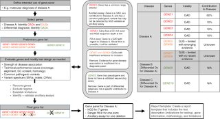
Gene sequencing panels are a powerful diagnostic tool for many clinical presentations associated with genetic disorders. Advances in DNA sequencing technology have made gene panels more economical, flexible, and efficient. Because the genes included on gene panels vary widely between laboratories in gene content (e.g., number, reason for inclusion, evidence level for gene–disease association) and technical completeness (e.g., depth of coverage), standards that address technical and clinical aspects of gene panels are needed. This document serves as a technical standard for laboratories designing, offering, and reporting gene panel testing.

Although these principles can apply to multiple indications for genetic testing, the primary focus is on diagnostic gene panels (as opposed to carrier screening or predictive testing) with emphasis on technical considerations for the specific genes being tested.

PANEL DESIGN CONSIDERATIONS

Phenotype-directed diagnostic gene panels Due to the sequencing capacity of current technologies, hundreds to thousands of genes can now be delved simultaneously to determine the cause of genetic disorders.

Examples of several phenotype-directed panels and subpanels are listed in Table 1. The goal of a diagnostic gene panel is to maximize clinical sensitivity and minimize the clinical burden from analysis of inappropriate or unnecessary genes that may result in variants of uncertain clinical significance (VUS). Patients may have limited opportunity for serial genetic tests, arguing for casting a wide diagnostic net; however, long lists of

(VUS), a possible outcome from testing large numbers of genes, can complicate medical management and cause unnecessary patient anxiety. When a panel is well-designed, it will

● Be cost-effective for a particular clinical indication.

● Maximize clinical sensitivity by, to the extent possible, including all GADs associated with a disorder, therebyallowing disorders with clinical heterogeneity and overlapping features to be molecularly diagnosed.

● Include GUSs with limited but emerging evidence ifadditional criteria are met (see example in “Clinicalsensitivity,” “Gene considerations,” and “Reporting”).

● Maximize clinical specificity by limiting or excludingGUSs, thereby minimizing detection of VUS.

● Employ auxiliary assays for genes/regions that cannot beinterrogated with current sequencing technology tomaximize the clinical utility.

Access Full PDF


Friday, January 8, 2021

Neuronavigation Systems: Concept, Techniques, Clinical Applications and Potential Pitfalls

Neuro-navigation, also called as frameless stereotactic surgery is the technique which involves performance of real-time intraoperative guidance during spinal or brain injuries. This increases safety and accuracy during neurosurgery. Neuro-navigation systems help to guide the surgeon to the surgical targets without the need for external frames. These systems are used majorly in brain surgeries which helps to limit the size of skull opening or craniotomy and remove brain lesions such as tumors or other tissue masses.

Download Sample PDF Of Neuronavigation Systems


There are significant technical issues that need to be addressed and understood in the use of such equipment in order to minimize inaccuracy. The most significant limitation of neuronavigation is brain shift. Brain shift can be minimized by selecting the site of the craniotomy uppermost in the operative field and by limited use of retraction, however, it is inevitable and can lead to limited application of the neuronavigation system over time during surgery. The loss of registration that can occur due to inadvertent intraoperative manipulation of the reference frame or loss of fixation of the head, can be retrieved by the registration of intraoperative bony landmarks at the margins of the craniotomy.

Localization and delineation of extent of lesions are critical for safe maximal resection of brain and spinal cord tumors. Neuronavigation systems have been developed for image-guided neurosurgery to aid in the accurate resection of brain tumors. Basic principles of navigated surgery are to see the tip of a pointer in an image space. A relationship between the device space and the image space has to be established. This operation is called registration or calibration of the navigation device. Basically, a transformation matrix (T) has to be calculated to map the coordinates of any point between the image and the device spaces. The aim of transformation matrix is to create a linkage between digital image data and anatomical structure, and therefore, to provide increasing 3-D orientation. Today's navigation systems provide approximately 2mm accuracy.

Stereoscopic navigation-controlled display of preoperative MRI and intraoperative 3D ultrasound is a new technology for minimally invasive image-guided surgery approaches in planning and guiding neurosurgery. Interactive stereoscopic visualization improves perception and enhances the ability to understand complex 3D anatomy. In 1947, Spiegel and Wycis performed the first stereotactic thalamotomy in humans, using the commissura posterior or pineal body as an internal individual reference system. Functional operations with similar frames and techniques were introduced by Talairach in Paris in 1949, by Riechert in Freiburg, Germany in 1952, and by Leksell in Stockholm in 1949 for the treatment of extrapyramidal movement disorders, intractable pain, epilepsy, and psychiatric disorders.

Advantages and disadvantages

There are some concerns about navigation systems including time consuming calculation and registration, restriction of space and view inside the operating field,and so on. Nevertheless, there are many advantages that can be helpful in the process of operation

Access Full PDF

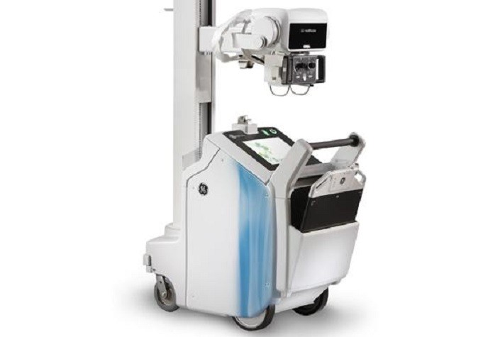
Mobile Radiography - Advanced Technology of Imaging

 Radiography is an imaging technique that used gamma rays, X-rays and other electromagnetic radiation to image the internal parts of a human...