Wednesday, November 25, 2020

What Is a Brain Pacemakers? How Pacemakers Treat Parkinson’s Disease?

 Electricity Restoring Function & Replacing Drugs

A brain pacemaker is a medical device implanted into the brain to stimulate the nervous tissues with electric signals. These pacemakers are being used widely to provide treatment to the patients having neurological disorders such as Parkinson's disease, epilepsy, and others. Other than giving stimulation to the brain, pacemakers also play an essential role in stimulating the spinal cord. Brain pacemakers have been found to offer a safe and effective procedure that provides symptomatic relief to patients. Doctor puts the pacemaker under the skin on your chest, just under your collarbone. It’s hooked up to your heart with tiny wires.

Download Sample PDF Brochure Of Study, Click Here


Doctors know that deep brain stimulation works as a therapy for Parkinson’s disease. But they’re still trying to figure out why and how. A new study sheds some light on the mechanism of action, suggesting that deep brain stimulation disrupts a pattern of excessively synchronized activity in the brain. In DBS, an implanted device sends tiny jolts of electricity through neurons, acting somewhat like a brain pacemaker. The technique is widely accepted as a treatment for Parkinson’s and other movement disorders; more than 100,000 patients have received implants that help control their tremors, rigidity, and other kinetic symptoms.

How does Pacemakers work?
• A pacemaker uses batteries to send electric signals to your heart to help it pump the right way.
• The pacemaker is connected to your heart by one or more wires. Tiny electric charges that you can’t feel move through the wire to your heart.
• Pacemakers work only when needed. They go on when your heartbeat is too slow, too fast or irregular.

The Brain Pacemaker has already revolutionized the treatment of movement disorders and in the future will completely alter the management of other disabling conditions. Our AIMS are to learn exactly how electrical stimulation normalizes brain function, to develop better technology for the device and other neural prosthetics, test these systems for new conditions, and thus improve patient’s quality of life.

Access Full PDF Brochure!

Friday, November 20, 2020

Quality Systems For Medical Imaging - Essentials for Physicians

Medical imaging is a process to examine the anatomical & physiological conditions of the human body for clinical analysis or diagnosis. It includes different processes to create image of internal organs & tissues for use in disease monitoring and treatment purposes. Medical imaging comprises three types of imaging- radiology imaging, nuclear imaging and optical imaging. Medical imaging seeks to reveal internal structures hidden by the skin and bones, as well as to diagnose and treat disease. Medical imaging also establishes a database of normal anatomy and physiology to make it possible to identify abnormalities. Although imaging of removed organs and tissues can be performed for medical reasons, such procedures are usually considered part of pathology instead of medical imaging.

Download Sample PDF Brochure Of Study


As a discipline and in its widest sense, it is part of biological imaging and incorporates radiology, which uses the imaging technologies of X-ray radiography, magnetic resonance imaging, ultrasound, endoscopy, elastography, tactile imaging, thermography, medical photography, and nuclear medicine functional imaging techniques as positron emission tomography (PET) and single-photon emission computed tomography (SPECT). Measurement and recording techniques that are not primarily designed to produce images, such as electroencephalography (EEG), magnetoencephalography (MEG), electrocardiography (ECG), and others, represent other technologies that produce data susceptible to representation as a parameter graph vs. time or maps that contain data about the measurement locations. In a limited comparison, these technologies can be considered forms of medical imaging in another discipline.

Medical imaging is often perceived to designate the set of techniques that noninvasively produce images of the internal aspect of the body. In this restricted sense, medical imaging can be seen as the solution of mathematical inverse problems. This means that cause (the properties of living tissue) is inferred from effect (the observed signal). In the case of medical ultrasound, the probe consists of ultrasonic pressure waves and echoes that go inside the tissue to show the internal structure. In the case of projectional radiography, the probe uses X-ray radiation, which is absorbed at different rates by different tissue types such as bone, muscle, and fat.

For More info in detail, Click here

Tuesday, November 17, 2020

Progress in Neuroengineering for Brain Repair | Future of Brain Implants, Potentially Safer way to Study

Intracerebral implants make use of drug-impregnated polymers that allow controlled release of the drug at the desired site in the brain over an extended period of time.

Brain implants are neural implants that are used to stimulate the parts & structures of the nervous system. These implants are technical systems that communicate with the nervous system and help to enhance senses, physical movement, and memory after a stroke or other head injuries. Deep brain stimulation and spinal cord stimulation are used to treat depression, obsessive-compulsive disorder and epilepsy, among other neural disorders.

Download Sample PDF Brochure of Brain Implants


This includes sensory substitution , e.g. in vision . Brain implants involve creating interfaces between neural systems and computer chips , popularly called brain-machine interfaces .Other brain implants are used in animal experiments simply to record brain activity for scientific reasons. Some brain implants involve creating interfaces between neural systems and computer chips. This work is part of a wider research field called brain-computer interfaces. (Brain-computer interface research also includes technology such as EEG arrays that allow interface between mind and machine but do not require direct implantation of a device.)

Neural implants such as deep brain stimulation and Vagus nerve stimulation are increasingly becoming routine for patients with Parkinson's disease and clinical depression, respectively. Next-generation implants may someday treat diseases of the brain itself, particularly those of aging. Such ailments are increasingly common as people live longer but devilishly hard to treat. One day, brain implants might “patch over” areas damaged by stroke or neurodegenerative illnesses, allowing healthy parts of the brain to communicate.

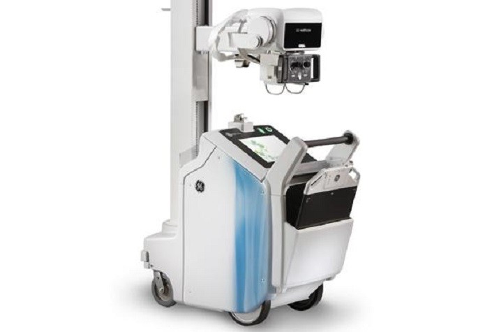
Mobile Radiography - Advanced Technology of Imaging

 Radiography is an imaging technique that used gamma rays, X-rays and other electromagnetic radiation to image the internal parts of a human...