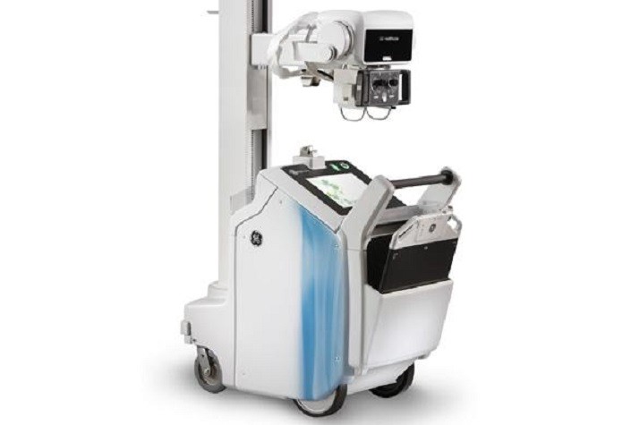
Medical imaging is a process to examine the anatomical & physiological conditions of the human body for clinical analysis or diagnosis. It includes different processes to create image of internal organs & tissues for use in disease monitoring and treatment purposes. Medical imaging comprises three types of imaging- radiology imaging, nuclear imaging and optical imaging. Medical imaging seeks to reveal internal structures hidden by the skin and bones, as well as to diagnose and treat disease. Medical imaging also establishes a database of normal anatomy and physiology to make it possible to identify abnormalities. Although imaging of removed organs and tissues can be performed for medical reasons, such procedures are usually considered part of pathology instead of medical imaging.
Download Sample PDF Brochure Of Study
As a discipline and in its widest sense, it is part of biological imaging and incorporates radiology, which uses the imaging technologies of X-ray radiography, magnetic resonance imaging, ultrasound, endoscopy, elastography, tactile imaging, thermography, medical photography, and nuclear medicine functional imaging techniques as positron emission tomography (PET) and single-photon emission computed tomography (SPECT). Measurement and recording techniques that are not primarily designed to produce images, such as electroencephalography (EEG), magnetoencephalography (MEG), electrocardiography (ECG), and others, represent other technologies that produce data susceptible to representation as a parameter graph vs. time or maps that contain data about the measurement locations. In a limited comparison, these technologies can be considered forms of medical imaging in another discipline.
Medical imaging is often perceived to designate the set of techniques that noninvasively produce images of the internal aspect of the body. In this restricted sense, medical imaging can be seen as the solution of mathematical inverse problems. This means that cause (the properties of living tissue) is inferred from effect (the observed signal). In the case of medical ultrasound, the probe consists of ultrasonic pressure waves and echoes that go inside the tissue to show the internal structure. In the case of projectional radiography, the probe uses X-ray radiation, which is absorbed at different rates by different tissue types such as bone, muscle, and fat.


No comments:
Post a Comment