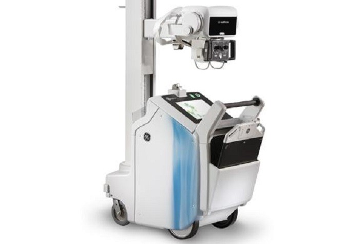
Friday, October 16, 2020

Advanced X-ray Detectors for Digital Radiography

X-ray detectors are used to measure varied properties of X-rays such as spatial distribution, flux, spectrum and others. With advancing technology and increasing demand for digital X-ray systems, the X-ray detectors require more robust structure with high transmission capability and temperature endurance, and resistant to ionizing radiations. X-rays consist of ionizing radiations, which are passed through the patient’s body and are absorbed by the internal organs. X-rays have been in use for non-invasive imaging of biological matters by passing high resolution radiations.

Download Sample PDF Brochure Of X-Ray Detectors


Digital radiography offers the potential of improved image quality as well as providing opportunities for advances in medical image management, computer-aided diagnosis and teleradiology. Image quality is intimately linked to the precise and accurate acquisition of information from the x-ray beam transmitted by the patient, i.e. to the performance of the x-ray detector. Detectors for digital radiography must meet the needs of the specific radiological procedure where they will be used. Key parameters are spatial resolution, uniformity of response, contrast sensitivity, dynamic range, acquisition speed and frame rate.

To obtain an image with any type of image detector the part of the patient to be X-rayed is placed between the X-ray source and the image receptor to produce a shadow of the internal structure of that particular part of the body. X-rays are partially blocked ("attenuated") by dense tissues such as bone, and pass more easily through soft tissues. Areas where the X-rays strike darken when developed, causing bones to appear lighter than the surrounding soft tissue.

Contrast compounds containing barium or iodine, which are radiopaque, can be ingested in the gastrointestinal tract (barium) or injected in the artery or veins to highlight these vessels. The contrast compounds have high atomic numbered elements in them that (like bone) essentially block the X-rays and hence the once hollow organ or vessel can be more readily seen. In the pursuit of nontoxic contrast materials, many types of high atomic number elements were evaluated. Unfortunately, some elements chosen proved to be harmful – for example, thorium was once used as a contrast medium (Thorotrast) – which turned out to be toxic, causing a very high incidence of cancer decades after use. Modern contrast material has improved and, while there is no way to determine who may have a sensitivity to the contrast, the incidence of serious allergic reactions is low.

For more info, Click Here!

Sunday, October 11, 2020

Principle of Neuroscience: Exploring the Science of Brain

Neuroscience is a study of that is concerned with the structure and function of the nervous system. The study covers the evolution, development, physiology, cellular & molecular biology, anatomy & pharmacology of the nervous system, and also behavioral, computational and cognitive neuroscience. Tools such as MRI scans and computerized 3-D models are used to perform tests for some common conditions including Down syndrome, Parkinson's disease, brain tumors, effects of stroke such as, language loss and many others.

Download Sample PDF Brochure Of Study, Click Here!


Humans have an estimated hundred billion neurons, or brain cells, each with about a thousand connections to other cells. One of the great challenges of modern neuroscience is to map out all the networks of cell-to-cell communication—the brain circuits that process all thoughts, feelings, and behaviors. The resulting picture, emerging bit by bit, is known as "the connectome." The ability of the brain to elaborate new connections and neuronal circuits—neuroplasticity—underlies all learning.

WHAT IS NEUROSCIENCE AND WHAT ARE ITS BRANCH SCIENCES?

In the case of humans, it is the branch of science that studies the brain, the spinal cord, the nerves extending from them, and the rest of the nervous systems including the synapses, etc. Recall that neurons, or nerve cells, are the biological cells that make up the nervous system, and the nervous system is the complex network of connections between those cells. In this connection, it may involve itself with the cellular and molecular bases of the nervous system as well as the systems responsible for sensory and motor activities of the body. It also deals with the physical bases of mental processes of all levels, including emotions and cognitive elements. Thus, it concerns itself with issues such as thoughts, mental activities, behaviors, the brain and the spinal cord, functions of nerves, neural disorders, etc. It wrestles with questions such as What is consciousness?, How and why do beings have mental activities?, What are the physical bases for the variety of neural and mental illnesses, etc.

A rapidly expanding discipline, neuroscience findings have grown by leaps and bounds over the past half-century. More work, however, will always be needed to fully understand the neural roots of human behavior, consciousness, and memory.

Access Full PDF Brochure

Friday, October 9, 2020

A Visual Approach to Diagnostic Imaging : Implementation & Monitoring

 Diagnostic imaging is well known across the globe with number of devices such as MRI, X-ray, CT scan and other devices used for viewing various structures within the body and determine presence of tumor in case of cancer. Diagnostic imaging devices have numerous advantages in the field of orthopedics, oncology, dentistry, and other major areas of medicine. These devices also monitor the disease condition thereby helping the healthcare provider to decide the line of treatment.


Diagnostic imaging describes a variety of non-invasive methods of looking inside the body to help determine the causes of an injury or an illness, and to confirm a diagnosis. It is also used to see how well your body is responding to a treatment for an illness or a fracture.

The type of imaging your doctor uses depends on your symptoms and the part of your body being examined. They include

  1. X-rays
  2. CT scans
  3. Nuclear medicine scans
  4. MRI scans
  5. Ultrasound

Many imaging tests are painless and easy. Some require you to stay still for a long time inside a machine. This can be uncomfortable. Certain tests involve exposure to a small amount of radiation. For some imaging tests, doctors insert a tiny camera attached to a long, thin tube into your body. This tool is called a scope. The doctor moves it through a body passageway or opening to see inside a particular organ, such as your heart, lungs, or colon. These procedures often require anesthesia.

Access Full PDF

Monday, October 5, 2020

Latest Advances in Neurorehabilitation Devices (Robots) : Future Challenges & Opportunity

Neurorehabilitation is a complex medical process to aid recovery from a nervous system injury, and the neurorehabilitation devices are used to the examination of the brain and the central nervous system and also providing solutions in the field of therapy and diagnoses.

Download Sample PDF Brochure Of Study, Click Here!

Neurorehabilitation Devices


The face of neurorehabilitation has progressively changed in recent years. Traditional neurorehabilitation procedures may have limited efficacy in most patients with common neurological diseases, such as stroke, Parkinson's disease, spinal cord injury, severe brain injury, spasticity, and cognitive disorders. New technologies have been reported to enhance the effectiveness of rehabilitation strategies in these conditions. They include robotic-assisted training, virtual reality, functional electrostimulation, non-invasive brain stimulation (NIBS) to enhance the intensity and quality of neurorehabilitation, and to manipulate brain excitability and plasticity, as well as innovative approaches such as assistive technology and domotics. The development of robotic devices for rehabilitation is a fast-growing field. Robotic rehabilitation is also widely used in the context of neurological disorders, where it is often provided in a variety of different fashions, depending on the specific function to be restored.

The development of rehabilitation robots started in the late 1980s. The following decade was a pioneering phase. After the year 2000, the first representatives of commercially available robots appeared. These devices can assist in practicing upper or lower limb movements and motor relearning, and in developing proprioception, cognitive functions, and attention. There is equipment that patients can use to practice the same movements as with the robots, but it does not provide mechanical assistance; so patients have to rely on their own strength. The emphasis is on high repetition, interactive and personalised therapy. The aim is to attain a higher level of function in a shorter time frame. The philosophy of the application of robots in rehabilitation is not to replace the therapist, but to widen treatment options.

The two main goals of therapy with rehabilitation robots are to develop upper limb function and to support gait re-education.

Access Full PDF Brochure

Friday, October 2, 2020

What is Medical Imaging Informatics? The Next Frontier in Healthcare Technology

 Medical imaging informatics (MII), aims to provide accurate, useful, and efficient interpretations of complicated images into simpler, communicable, and in a useful manner. It mainly comprises of the application, development, and assessment of information that are useful to treat patients effectively.

Download PDF Brochure Of Study, Click Here


There are rapid changes occurring in the health care environment. Radiologists face new challenges but also new opportunities. The purpose of this report is to review how new informatics tools and developments can help the radiologist respond to the drive for safety, quality, and efficiency. These tools will be of assistance in conducting research and education. They not only provide greater efficiency in traditional operations but also open new pathways for the delivery of new services and imaging technologies. Our future as a specialty is dependent on integrating these informatics solutions into our daily practice.

Imaging informatics exists at the intersection of several broad fields:

  1. Biological science — includes bench sciences such as biochemistry, microbiology, physiology and genetics
  2. Clinical services — includes the practice of medicine, bedside research, including outcomes and cost-effectiveness studies, and public health policy
  3. Information science — deals with the acquisition, retrieval, cataloging, and archiving of information
  4. Medical physics / biomedical engineering — entails the use of equipment and technology for a medical purpose
  5. Cognitive science — studying human computer interactions, usability, and information visualization
  6. Computer science — studying the use of computer algorithms for applications such as computer assisted diagnosis and computer vision

This article discusses basic imaging informatics protocols, picture archiving and communication systems, and the electronic medical record. It details key instrumentation and data mining technologies used in medical imaging informatics as well as practical operational issues, such as procurement, maintenance, teleradiology, and ethics. The technologies of medical imaging and radiation therapy are so complex and computer-driven that it is difficult for physicians and technologists responsible for their clinical use to know exactly what is happening at the point of care. Medical physicists are best equipped to understand the technologies and their applications, and these individuals are assuming greater responsibilities in the clinical arena to ensure that intended care is delivered in a safe and effective manner. Built on a foundation of classic and cutting-edge research, Informatics in Medical Imaging supports and updates medical physicists functioning at the intersection of radiology and radiation.

Access Full PDF

Mobile Radiography - Advanced Technology of Imaging

 Radiography is an imaging technique that used gamma rays, X-rays and other electromagnetic radiation to image the internal parts of a human...